Female Internal Reproductive Organs Anatomy - Female Internal Organ Diagram . Female Internal Organ ... - This pathway consists of the following. Reproductive physiology lectures female reproductive anatomy. Find stockbilleder af female internal reproductive organs anatomy3d i hd og millionvis af andre royaltyfri stockbilleder, illustrationer og vektorer i shutterstocks samling. The uterus and ovaries are particularly affected by atrophy (shrinkage) after the menopause. — written by rachel nall, msn, crna on november 5, 2019. ♦ fibrous, collagenous organ with a small amount of muscle.
♦ fibrous, collagenous organ with a small amount of muscle. It is a fibromuscular canal lined with stratified squamous epithelium that leads from the uterus to the vulva. External organs and internal organs of the female reproductive system with structure, functions and diagram. The area containing these organs is called the vulva. This pathway consists of the following
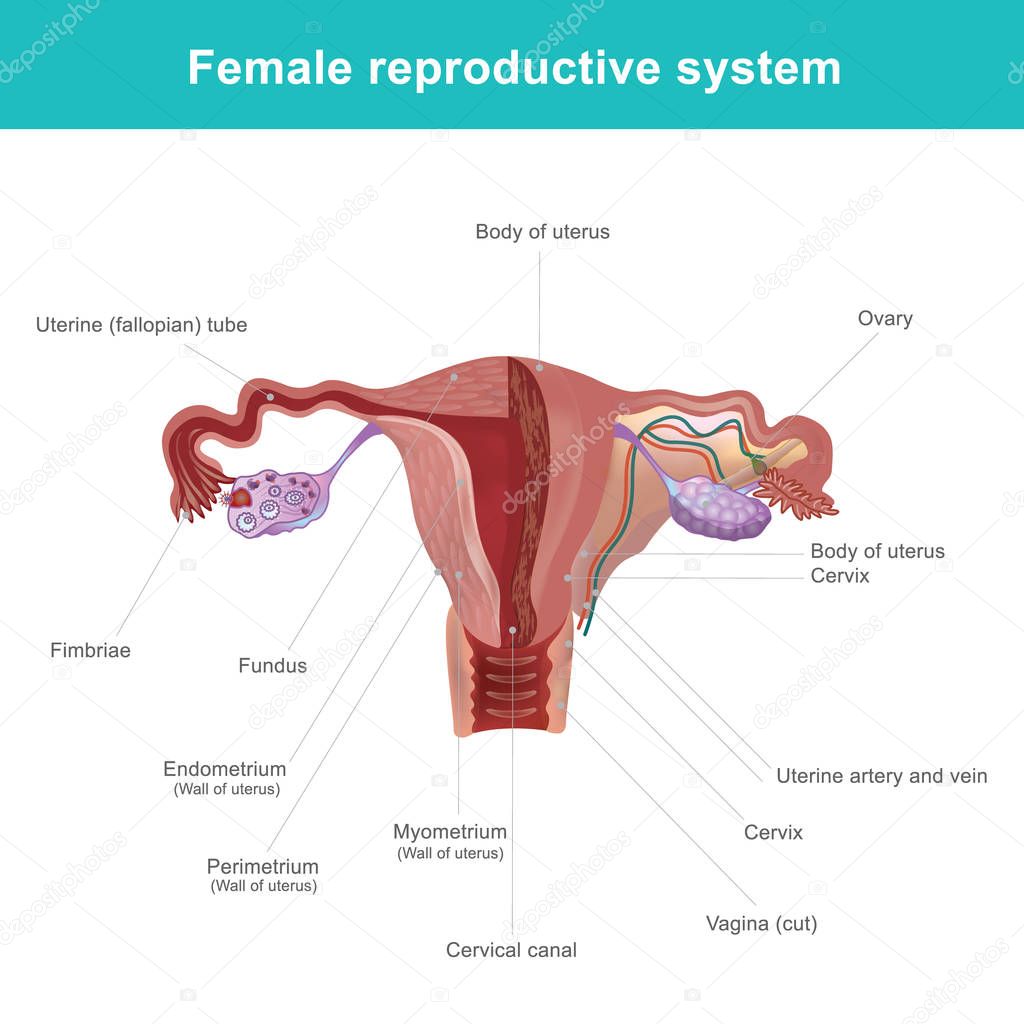
Female reproductive organs (Anatomy) - Study Guide | Kenhub from thumbor.kenhub.com It is a fibromuscular canal lined with stratified squamous epithelium that leads from the uterus to the vulva. The female reproductive system includes the ovaries, fallopian tubes, uterus, vagina, vulva, mammary glands and breasts. The function of the external female reproductive structures (the genitals) is twofold: • uterine artery supplies the rest of the uterine horns and the uterine body. External organs and internal organs of the female reproductive system with structure, functions and diagram. Corresponds to the level of the internal os of the uterus. Tusindvis af nye billeder af høj kvalitet tilføjes hver dag. Introduction • the reproductive organ in female are those which concerned with copulation, fertilization, growth and development of fetus and its subsequent exit to the outer world.
Together they comprise the female reproductive system, supporting sexual female reproductive organs undergo substantial structural and functional changes every month.
• uterine artery supplies the rest of the uterine horns and the uterine body. The site of the histological internal os is where the mucous membrane of the isthmus becomes that of the cervix. A thin membrane of tissue. Female internal reproductive organs anatomy. Find more on the female reproductive organs, the menstrual cycle, and more. External structures include the mons pubis, pudendal the female reproductive system contains two main parts: Most common malignancy of the female reproductive tract. Female reproductive system, human internal organ anatomy vector illustration on a white background. The area containing these organs is called the vulva. Introduction • the reproductive organ in female are those which concerned with copulation, fertilization, growth and development of fetus and its subsequent exit to the outer world. This is a branch off the internal iliac artery in most domestic species, except the mare where instead it is a. It is a fibromuscular canal lined with stratified squamous epithelium that leads from the uterus to the vulva. The function of the external female reproductive structures (the genital) is twofold:
Most common malignancy of the female reproductive tract. Female woman android with internal technology vector. — written by rachel nall, msn, crna on november 5, 2019. The site of the histological internal os is where the mucous membrane of the isthmus becomes that of the cervix. The function of the external female reproductive structures (the genitals) is twofold:
The female reproductive system. — Stock Vector ... from st3.depositphotos.com A thin membrane of tissue. These changes are not only there to make women's. The area containing these organs is called the vulva. This pathway consists of the following The site of the histological internal os is where the mucous membrane of the isthmus becomes that of the cervix. Female woman android with internal technology vector. External structures include the mons pubis, pudendal the female reproductive system contains two main parts: Female organs, cells and structures.
• uterine artery supplies the rest of the uterine horns and the uterine body.
Our experts describe the functions of female reproduction, including ovulation, fertilization, and menopause. • uterine artery supplies the rest of the uterine horns and the uterine body. Medically reviewed by carolyn kay, m.d. Female woman android with internal technology vector. Most common malignancy of the female reproductive tract. The internal reproductive organs vagina: The function of the external female reproductive structures (the genitals) is twofold: Female reproductive anatomy and physiology. This is a branch off the internal iliac artery in most domestic species, except the mare where instead it is a. This pathway consists of the following The area containing these organs is called the vulva. Biology of the female reproductive system. External organs and internal organs of the female reproductive system with structure, functions and diagram.
The function of the external female reproductive structures (the genital) is twofold: Tusindvis af nye billeder af høj kvalitet tilføjes hver dag. Learn about the female reproductive system's anatomy through diagrams and detailed facts. Our experts describe the functions of female reproduction, including ovulation, fertilization, and menopause. This article looks at female body parts and their functions, and it a guide to female anatomy.
Labeled Female Anatomical Diagram : Male And Female ... from images.medicaldaily.com A thin membrane of tissue. Which of the following is not an access… female reproductive anatomy. Introduction • the reproductive organ in female are those which concerned with copulation, fertilization, growth and development of fetus and its subsequent exit to the outer world. The uterus consists of three layers: Solid, ovoid structures located within the. The female reproductive anatomy includes parts inside and outside the body. — written by rachel nall, msn, crna on november 5, 2019. Female organs, cells and structures.
Tusindvis af nye billeder af høj kvalitet tilføjes hver dag.
These changes are not only there to make women's. Female organs, cells and structures. This pathway consists of the following • uterine artery supplies the rest of the uterine horns and the uterine body. The function of the external female reproductive structures (the genitals) is twofold: The area containing these organs is called the vulva. An female's internal reproductive organs are the vagina, uterus, fallopian tubes, cervix, and ovary. Female reproductive system, human internal organ anatomy vector illustration on a white background. External organs and internal organs of the female reproductive system with structure, functions and diagram. ♦ fibrous, collagenous organ with a small amount of muscle. — written by rachel nall, msn, crna on november 5, 2019. The internal reproductive organs vagina: Introduction • the reproductive organ in female are those which concerned with copulation, fertilization, growth and development of fetus and its subsequent exit to the outer world.
Female anatomy includes the external genitals, or the vulva, and the internal reproductive organs female internal. • uterine artery supplies the rest of the uterine horns and the uterine body.

:fill(FFFFFF,true):format(jpeg)/images/container/female-reproductive-organs/Female_reproductive_organs_1.png)


0 Comments